What Do Breast Masses Look Like on a Mammogram?
What shows up on your mammogram, and what does it look like? See what benign and malignant masses look like on a mammogram. Mammograms help with early detection and screening for breast cancer. You can see areas of dark and light, which correspond to normal and dense breast tissue. Breast masses will appear light (whiter) because they are denser than other features in the breast. Images are of actual mammograms, courtesy of the National Cancer Institute.
Please note: Red arrows were added to help you see the part of the image that is being featured.
Normal Fatty Breast Tissue on a Mammogram
Dark areas are fatty tissue; light areas are denser tissue which contain ducts, lobes, and other findings.

Description: Shown is a mammogram of a normal fatty breast, typical of older women. Diagnosis of abnormal lesions or cancer is more accurate in non-dense breasts.
Mammograms work best on fatty breasts because they have less areas of density (whiter masses). Breast masses which usually cause concern are lighter than normal dense tissue.
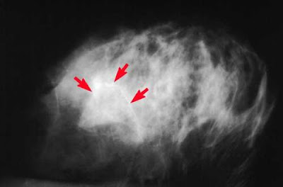
Breast Calcifications on a Mammogram
Dark areas are normal fatty breast tissue. Lighter areas are denser tissue. The whiter spots are calcifications.

Description: This abnormal mammogram is not necessarily cancerous. Also seen are calcifications through ductal patterns. The patient would have a follow-up mammogram in three months for a comparison.
Microcalcifications are tiny bits of calcium that may show up in clusters or in patterns (like circles) and are associated with extra cell activity in breast tissue. Usually the extra cell growth is not cancerous, but sometimes tight clusters of microcalcifications can indicate early breast cancer. Scattered microcalcifications are usually a sign of benign breast tissue.
Breast Tumor on a Mammogram
Dark areas are normal fatty breast tissue. Lighter areas are denser tissue. The whitest area is the most dense, indicating a tumor (breast cancer).

Description: Shown is a mammogram of a fatty breast with an obvious cancer, indicated by an arrow in lower right corner.
A cancerous tumor in the breast is a mass of breast tissue that is growing in an abnormal, uncontrolled way. The tumor may invade surrounding tissue or shed cells into the bloodstream or lymph system.
What shows up on your mammogram, and what does it look like? See what benign and malignant masses look like on a mammogram. Mammograms help with early detection and screening for breast cancer. You can see areas of dark and light, which correspond to normal and dense breast tissue. Breast masses will appear light (whiter) because they are denser than other features in the breast. Images are of actual mammograms, courtesy of the National Cancer Institute.
Please note: Red arrows were added to help you see the part of the image that is being featured.
Normal Fatty Breast Tissue on a Mammogram
Dark areas are fatty tissue; light areas are denser tissue which contain ducts, lobes, and other findings.

Description: Shown is a mammogram of a normal fatty breast, typical of older women. Diagnosis of abnormal lesions or cancer is more accurate in non-dense breasts.
Mammograms work best on fatty breasts because they have less areas of density (whiter masses). Breast masses which usually cause concern are lighter than normal dense tissue.
Breast Calcifications on a Mammogram
Dark areas are normal fatty breast tissue. Lighter areas are denser tissue. The whiter spots are calcifications.

Description: This abnormal mammogram is not necessarily cancerous. Also seen are calcifications through ductal patterns. The patient would have a follow-up mammogram in three months for a comparison.
Microcalcifications are tiny bits of calcium that may show up in clusters or in patterns (like circles) and are associated with extra cell activity in breast tissue. Usually the extra cell growth is not cancerous, but sometimes tight clusters of microcalcifications can indicate early breast cancer. Scattered microcalcifications are usually a sign of benign breast tissue.
Breast Tumor on a Mammogram
Dark areas are normal fatty breast tissue. Lighter areas are denser tissue. The whitest area is the most dense, indicating a tumor (breast cancer).

Description: Shown is a mammogram of a fatty breast with an obvious cancer, indicated by an arrow in lower right corner.
A cancerous tumor in the breast is a mass of breast tissue that is growing in an abnormal, uncontrolled way. The tumor may invade surrounding tissue or shed cells into the bloodstream or lymph system.
No comments:
Post a Comment